Current Issue: Volume 1 | Issue 2 | Year 2025
Institutional Subscription
Individual Subscription
Case Reports
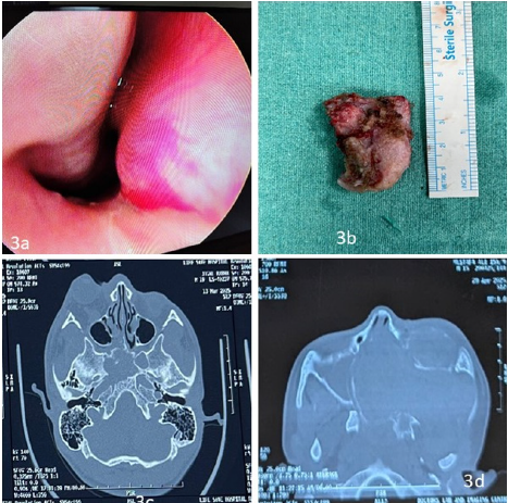
A Rare Case of Unusually Big Adenomatoid Odontogenic Tumor
Sounak Patra, Manu CB, Sauradeep Das, Suvamoy Chakraborty
Abstract
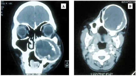
Of all odontogenic tumors, adenomatoid odontogenic tumors (AOT) are extremely rare, making for about 3% of cases. It is a benign, slowly developing, encapsulated, non-invasive, and non-aggressive odontogenic lesion connected to an impacted tooth. For years, these lesions could go undetected. The lesion does not return after the standard course of treatment of enucleation and curettage.1
Here, we report an uncommon case of an unusually large AOT in a 16-year-old female patient from northeastern India, who presented with complaints of a swelling over her left side of face for past 5 years associated with bilateral nasal blockage. Clinical examination and investigations revealed a hard swelling with some cystic regions of size 7.6 (CC) x 7.2 (AP) x 6.6 (TR) cm. The patient underwent staged removal of the tumor and the histopathological examination of the cyst wall showed features suggestive of adenomatoid odontogenic tumor. The patient remains symptom free on a follow up period of 18 months.
Keywords: Adenomatoid Tumor, Odontogenic Cyst, Odontodysplasia
Abstract
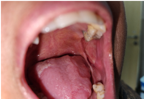
Introduction: Angina bullosa hemorrhagic is a benign, rare condition of oral cavity, characterized by a sudden onset blood filled blisters in mucosal of oral cavity which rupture and heals spontaneously without scarring, considered to present in 0.05% of cases of oral ulcer. It resolves spontaneously, but in 30% cases it has tendency to reoccur.
Case report: A 48-year-old female presented with sudden onset, self-resolving oral ulcer clinically diagnosed as Angina Bullosa Hemorrhagic following Ordioni et al. criteria for diagnosis.
Conclusion: Angina Bullosa Hemorrhagic is a rare, benign disease of oral cavity that rupture and heals spontaneously without scarring, often triggered by trauma or associated with systemic factors such as hypertension, diabetes or prolonged use of corticosteroid. Diagnosis is mainly clinical. Differentiation form vesicobullous, hematological disorder is essential to avoid misdiagnosis and unnecessary interventions.
Angiomyolipoma of the Parotid Gland: A Rare Presentation
Anupam Mohanty, Sauradeep, Suvamoy Chakraborty, Manu, Biswajit
Summary
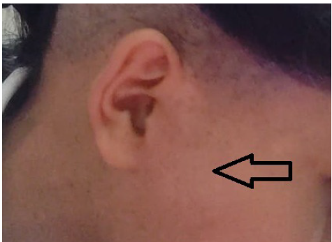
We present the case of a girl in her late teens who presented with a slowly enlarging, painless mass in her
right parotid gland over four years. Initial investigations, including ultrasonography and MRI, suggested a vascular malformation. Fine needle aspiration supported a benign tumour. Histological analysis following excision confirmed the diagnosis of angiomyolipoma (AML) of the parotid gland. This case highlights the importance of considering rare benign mesenchymal tumours like AML in the differential diagnosis of parotid gland masses, even in the absence of typical associations such as tuberous sclerosis.
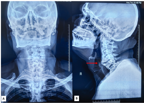
Cervical Osteophyte-Induced Dysphagia: A Case Report
Nongthombam Bidyananda Singh, Manu CB, Suvamoy Chakraborty
Summary
We present the case of a girl in her late teens who presented with a slowly enlarging, painless mass in her
right parotid gland over four years. Initial investigations, including ultrasonography and MRI, suggested a vascular malformation. Fine needle aspiration supported a benign tumour. Histological analysis following excision confirmed the diagnosis of angiomyolipoma (AML) of the parotid gland. This case highlights the importance of considering rare benign mesenchymal tumours like AML in the differential diagnosis of parotid gland masses, even in the absence of typical associations such as tuberous sclerosis.
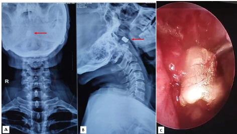
Complex Retained Ballistic Foreign Bodies of the Nasopharynx and Ethmoid Sinus: A Two-Case Series and Review
Nongthombam Bidyananda Singh, Sauradeep Das, Manu CB, Suvamoy Chakraborty
Abstract
Background: Retained ballistic foreign bodies in anatomically intricate regions such as the nasopharynx or paranasal sinuses are exceptionally rare and pose diagnostic and therapeutic challenges.
Cases: We present two cases in 49-year-old males: Case 1 involved a bullet lodged in the posterior nasopharyngeal wall with multiple tooth fragments embedded in the tongue following intraoral gunshot trauma; Case 2 featured a metallic fragment retained in the left posterior ethmoid sinus after a self-inflicted nasal gunshot wound.
Management and Outcome: Both patients underwent emergency surgical intervention—open retrieval and tracheostomy for Case 1, endoscopic sinus surgery with septoplasty for Case 2—and recovered uneventfully.
Conclusion: These cases underscore the critical role of high resolution computed tomography, multidisciplinary planning, and tailored surgical approaches in successfully managing rare upper aerodigestive ballistic injuries.
Keywords: Ballistic trauma; retained foreign body; nasopharynx; ethmoid sinus; endoscopic sinus surgery; Computed tomography scan imaging; gunshot wound
Case Series
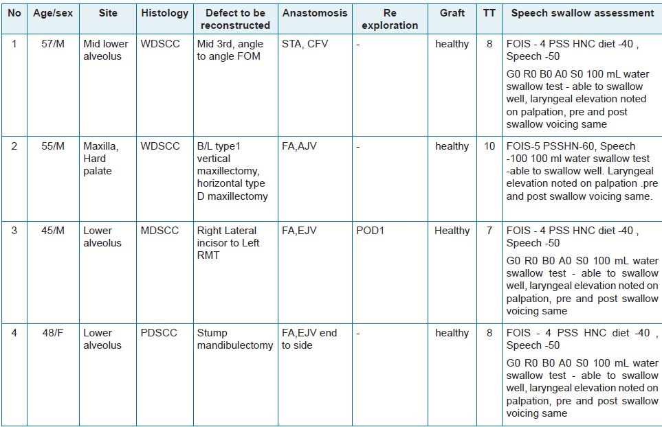
Yamuna Ranganathan1, Siddhartha Basuroy2
Abstract
Head and neck cancers are managed by a multimodality approach. However, after oncologic resection, reconstruction should focus on restoring form and function. Herein, we report a series of 04 cases who underwent resection of oral cavity tumors followed by reconstruction with a free fibula osteocutaneous flap and its functional outcome.
Keywords: Free fibula osteocutaneous flap, reconstruction, functional outcome
Original Articles
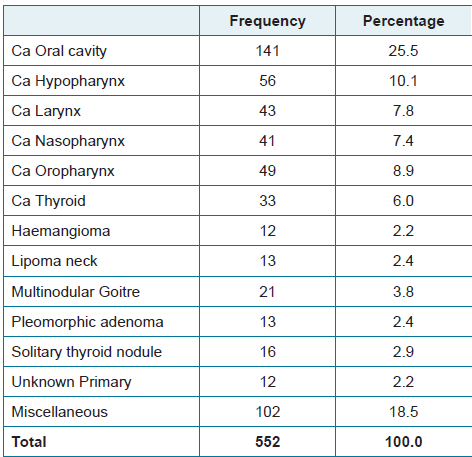
Demographic Profile of Head and Neck Malignancy in Northeast India:
A Retrospective Study
Rahul RC, Neizekhutuo Brian Shunyu, Gautam Sharma, Partha Pratim Medhi, Jijitha Lakshmanan, Ratan Medhi, Taniyang Lailyang
Abstract
Introduction: Head and neck cancers (HNCs) are the sixth most common malignancy worldwide, with 57.5% of the global burden occurring in Asia, particularly in India, particularly in the north-eastern region where genetic, environmental and lifestyle-related risk factors are significant contributors.
Methods: A retrospective study was carried out, collecting data from Hospital base’s cancer clinic registry for the period of January 2024–June 2025.
Results: Total of 552 patient data were collected, among them males are more prevalent than female, with elder people of age 45–60 years of age were more affected by cancer. The majority of patients were from Assam (78.6%), followed by Nagaland (16.8%), with sporadic cases from other states of the North east. The most common malignancy was found to be oral cavity carcinoma (25.5%), followed by hypopharyngeal carcinoma (10.1%), oropharyngeal carcinoma (8.9%), laryngeal carcinoma (7.8%), and nasopharyngeal carcinoma (7.4%). Among benign or non-malignant lesions, multinodular goitre (3.8%), lipoma (2.3%), and pleomorphic adenoma (2.3%) are predominated. Miscellaneous tumors accounted for 18.5%.
Discussion: This one and half year study highlights that head and neck cancers in North-East India mostly affect males in their middle to late adulthood, with oral cavity carcinoma being the most prevalent subtype. Assam and Nagaland contribute the highest case burden.
Conclusion: High number of cases, with wide verities of malignancies rises concern and needs targeted awareness, early screening initiatives, and region-specific cancer control
Epistaxis: Patterns of Presentation and Management – A Retrospective
Study from A Tertiary Institution
Aswathi Kallyadan Veetil, Jijitha Lakshmanan, Neizekhotuo Brian Shunyu, Hanifa Akthar, Ruuzeno Kuotsu, Nisha Kumari, Paramesh Patra
Abstract
Introduction: Epistaxis is a common ENT emergency, with varied presentations ranging from minor nasal bleeding to life-threatening hemorrhage. The aim of the study was to evaluate the varying presentations, etiologies, and management outcomes of epistaxis in a tertiary care center in Northeast India.
Methods: Retrospective review of the details of the patients admitted with epistaxis from April 2024–April 2025 at a tertiary hospital in Northeast India. Data from 114 patients presenting with epistaxis were analyzed for demographics, bleeding site, etiology, comorbidities, and treatment methods.
Results: Of 114 patients, 62.2% were male, with mean age of 41.3 +17.2 years. Anterior epistaxis accounted for 95.5% of cases. The leading causes were hypertension (35.8%) and nasal trauma (20.9%). Most cases were managed conservatively with nasal compression, topical agents, and cautery. Anterior nasal packing was done in 35.8% of patients, while 6 patients required surgical or radiological intervention.
Conclusion: Anterior epistaxis was the most frequent presentation, primarily due to hypertension and trauma. Conservative measures were effective in most cases. Surgical or interventional procedures were reserved for refractory cases, supporting a stepwise management approach.